Fall 2026 (Volume 36, Number 3)
Deciphering Ophthalmology Referrals and Assessments for Shared Care: A Practical Guide for Rheumatologists
By Alessana Carmona, MD; Brian J. Chan, MD, FRCSC; and Raj Carmona, MBBS, FRCPC
Download PDF
Introduction
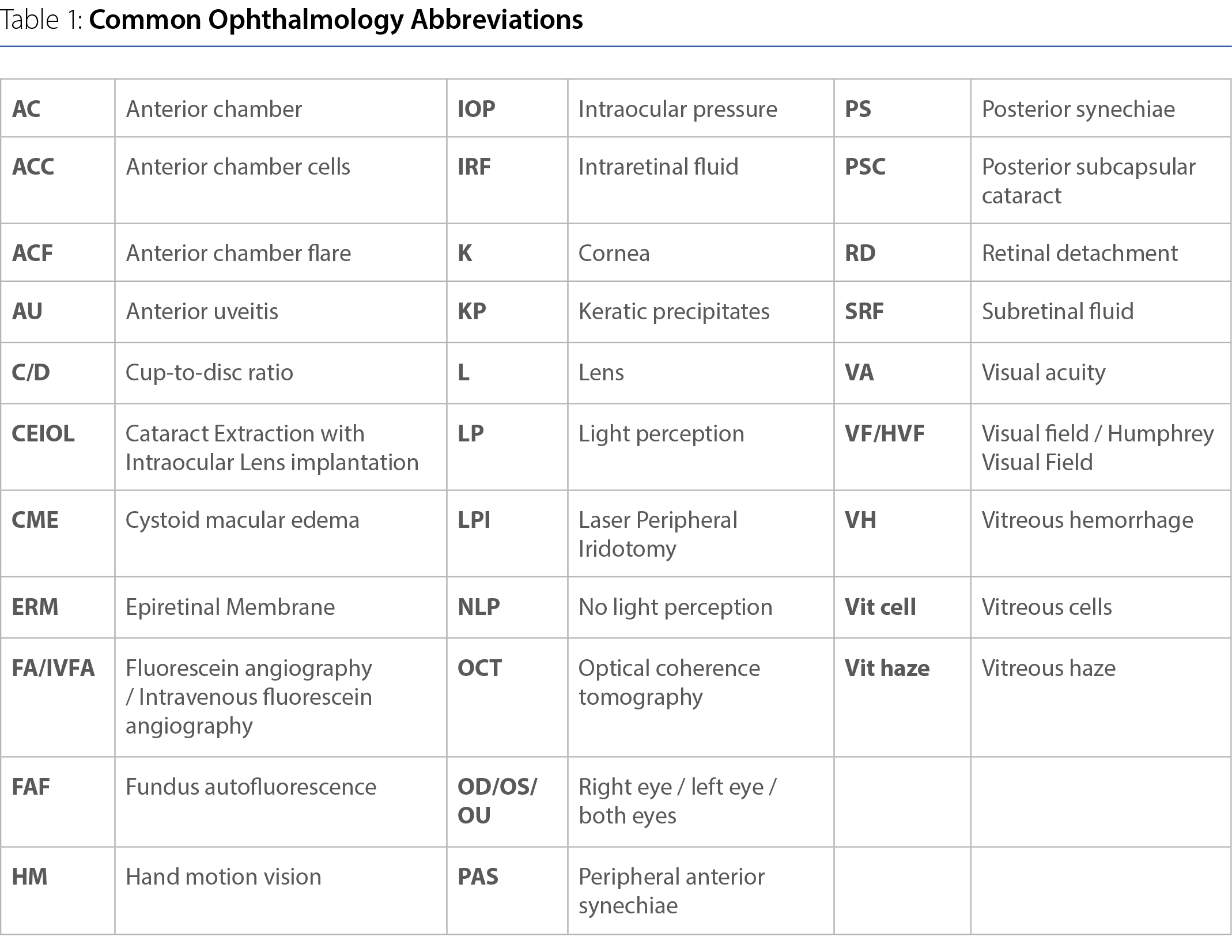
As experts in inflammatory/autoimmune diseases and immunotherapy, rheumatologists are uniquely equipped to co-manage patients with inflammatory ocular diseases (IOD), both primary ocular disorders and those associated with systemic diseases. However, due to extensive use of abbreviations (Table 1) and short forms, as well as ocular investigations and assessments of disease activity that are less familiar to rheumatologists, interpreting ophthalmology referrals and ocular assessments can be like deciphering code. This article serves to crack that code and improve shared care of IOD patients.
Anatomic Involvement and Differential Diagnoses
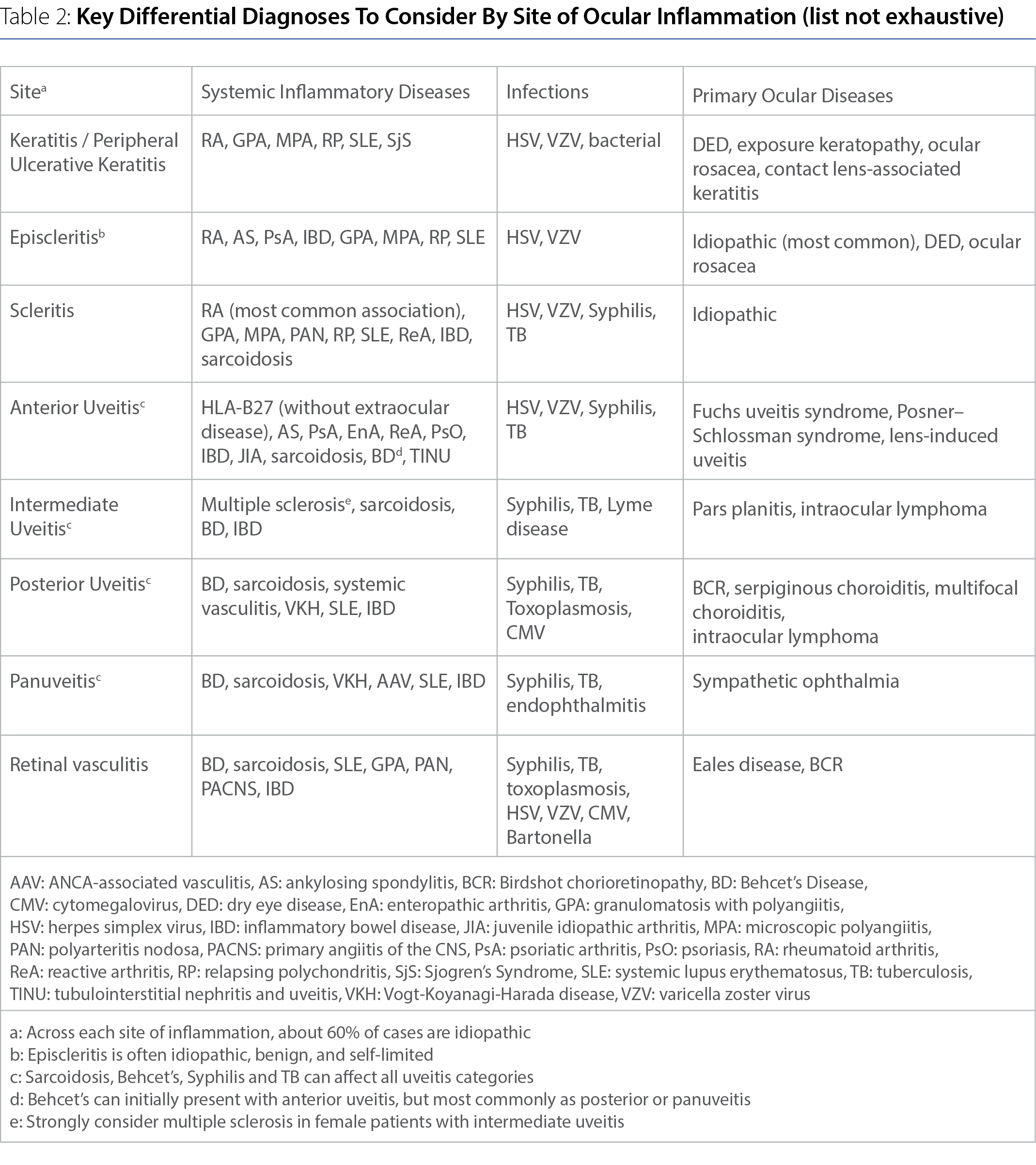
Familiarity with ocular anatomy (Figure 1) aids interpretation of ophthalmic findings. The anterior segment includes the cornea, aqueous humor, iris, ciliary body, lens, anterior chamber (between the cornea and iris), and posterior chamber (between the iris and lens). The posterior segment includes the vitreous body, retina, choroid and optic nerve. The Standardization of Uveitis Nomenclature (SUN) Working Group classifies uveitis as anterior (iris +/- ciliary body), intermediate (vitreous and pars plana), posterior (retina and/or choroid), and panuveitis (entire uveal tract).1 Referrals should specify the site of inflammation and anatomic extent, as this guides the differential diagnosis (Table 2).
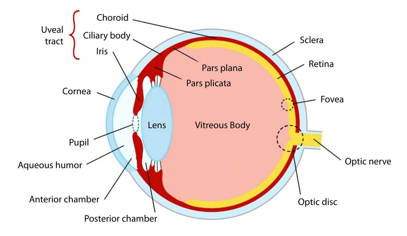
Figure 1: Ocular Anatomy Cross-section
Illustration by Zachary Carmona, 2026.
Anterior Segment Findings on Slit Lamp Examination
Key slit lamp findings in anterior uveitis include ciliary injection, anterior chamber cells (leucocytes suspended in aqueous humor), flare (protein leakage causing a smoky appearance), keratic precipitates (inflammatory deposits on the corneal endothelium), posterior synechiae (iris adhesions onto the lens), hypopyon (pooled leucocytes in the anterior chamber), iris nodules, and cataract formation. Anterior chamber cells are graded using a 1 mm ×
1 mm slit beam as: 0 (<1 cell), 0.5+ (1–5 cells), 1+ (6–15), 2+ (16–25), 3+
(26–50), and 4+ (>50 cells). 1+ or higher is considered active inflammation. Flare is graded 0 to 4+ according to increasing protein haze and impaired visualization of iris/lens details.1
Importantly, “granulomatous” uveitis refers to the clinical appearance of large “mutton-fat” keratic precipitates and iris nodules, rather than pathologic granulomas, and is associated with conditions such as sarcoidosis, tuberculosis, herpetic infections, syphilis, and Vogt-Koyanagi-Harada disease (VKH). Uveitis associated with HLA-B27-associated diseases is usually non-granulomatous, but exceptions occur.
Posterior Segment Findings on Slit Lamp Examination and Indirect Ophthalmoscopy
Despite being anterior in location, anterior uveitis may be associated with “spillover” anterior vitritis, and cystoid macular edema (CME).
Intermediate uveitis is characterized by vitreous cells, snowballs (inflammatory aggregates), and snowbanking over the pars plana. Inflammatory vitreous haze is graded as: 0 (clear), 0.5+ (trace), 1+ (mild blurring of retinal details), 2+ (moderate blurring, vessels still visible), 3+ (severe, only large vessels visible) and 4+ (very severe, fundus barely visible).1
Posterior uveitis may present with retinitis, choroiditis, chorioretinitis, retinal vasculitis, optic disc edema, and macular edema. Retinal vasculitis can manifest as vascular sheathing (coated appearance), hemorrhage, ischemia, and cotton wool spots. Chronic inflammation may lead to epiretinal membrane formation, retinal neovascularization, and optic atrophy.
Visual Acuity (VA) Assessment
VA is typically measured using a Snellen chart at 20 feet. 20/20 is normal visual acuity. 20/40 means the patient sees at 20 feet what a normal observer sees at 40 feet.2 20/200 is the threshold for legal blindness.3
Acute VA loss may result from corneal edema, active anterior chamber inflammation, vitritis, macular edema, retinal ischemia, or optic nerve involvement. Chronic reductions may indicate cataract, glaucoma, retinal scarring, or macular atrophy.
Visual Field (VF) Assessment
Automated VF testing evaluates retinal sensitivity across the VF, often using Humphrey automated perimetry. The patient fixates on a central target while lights of varying intensities are presented at different retinal locations.
VF assessment is essential in glaucoma, optic neuropathy, retinal disease, and hydroxychloroquine screening. Hydroxychloroquine retinopathy classically produces paracentral scotomas on 10-2 VF testing, although Asian patients may demonstrate more peripheral defects on 24-2 or 30-2 testing.4 Mild nonspecific defects are common and do not necessarily indicate retinal toxicity.
Intraocular Pressure (IOP)
IOP is usually measured using applanation tonometry. Normal IOP is generally 10–21 mmHg.2
Acute inflammation can decrease IOP due to ciliary body dysfunction, whereas chronic inflammation can increase IOP secondary to trabecular meshwork damage or synechiae. Topical, intravitreal/periocular, and systemic corticosteroids can increase IOP, potentially leading to secondary glaucoma. Importantly, the ophthalmologic term “steroid response” refers to corticosteroid-induced (usually topical) elevation of IOP, rather than a therapeutic benefit.3
Optical Coherence Tomography (OCT)
OCT is a non-invasive imaging modality that produces high-resolution cross-sectional images of the retina using reflected light waves.
OCT can detect macular pathology that may not be visible clinically, including CME. CME is a critical finding as it can lead to vision loss. OCT may also demonstrate epiretinal membranes, subretinal fluid, vitreomacular traction, retinal atrophy, and disruption of photoreceptor layers. Serial OCT imaging is frequently used to monitor therapeutic response.3
Intravenous Fluorescein Angiography (IVFA)
IVFA involves intravenous injection of sodium fluorescein dye followed by serial retinal photography. The early arterial phase assesses retinal and choroidal perfusion, the venous phase evaluates vascular filling, and late frames identify leakage, staining, or ischemia.3
In choroiditis and chorioretinitis, active lesions are often hypofluorescent early and hyperfluorescent late due to staining and leakage. Retinal vasculitis produces vascular leakage, capillary non-perfusion, and optic disc leakage.
Behçet’s disease classically demonstrates diffuse retinal vascular leakage, capillary dropout, and occlusive vasculitis.5 Sarcoidosis commonly causes segmental venous leakage and periphlebitis (“candle-wax drippings”).6 VKH demonstrates multiple pinpoint leaks with pooling in serous retinal detachments.7 ANCA-associated vasculitis may produce retinal ischemia, vascular occlusion, and optic nerve leakage.8
Importantly, “retinal vasculitis” in ophthalmology is defined primarily by clinical and angiographic features such as vascular leakage, staining, sheathing, and non-perfusion, rather than the histopathologic vessel wall inflammation and necrosis seen in systemic vasculitis. Retinal vascular inflammation in infectious or immune-mediated posterior uveitis does not, in isolation, indicate a primary retinal vasculitis syndrome or systemic necrotizing vasculitis.
Topical Therapies for Non-infectious Anterior Uveitis
For non-infectious anterior uveitis, first-line therapy is usually a topical corticosteroid (e.g. prednisolone acetate 1%). Dosing is initially intensive (e.g. hourly) with gradual taper over 4-6 weeks. Adjunctive cycloplegic/mydriatic agents (e.g., cyclopentolate 1%) relieve pain by reducing ciliary muscle spasm and help prevent posterior synechiae by dilating the iris away from the lens.3 There is usually no role for topical therapies in posterior segment involvement.
Indications for Systemic Immunotherapy in Non-Infectious Uveitis
Systemic immunotherapy is indicated for recurrent anterior uveitis, failure of local therapy or steroid intolerance, bilateral disease, posterior segment involvement, chronic uveitis, or any vision-threatening disease.
Systemic corticosteroids (e.g. prednisone 0.5–1 mg/kg/day or equivalent) are used for rapid suppression of active inflammation. Intravenous methylprednisolone is used in some cases of severe posterior or panuveitis, or retinal vasculitis.9
Conventional steroid-sparing agents include methotrexate, mycophenolate mofetil, azathioprine, and cyclosporine. Biologic therapy, particularly adalimumab or infliximab, is widely used in refractory non-infectious uveitis.10
Conclusion
Disease activity and treatment response are assessed by slit-lamp assessment of AC cells/flare, vitreous haze and retinal/choroidal lesions, OCT evidence of CME or serous detachment, IVFA leakage and non-perfusion, functional measures such as VA and VF, and topical corticosteroid requirements. Understanding ophthalmology terminology and assessment enables rheumatologists to participate more effectively in shared care of IOD patients, with timely escalation of systemic immunotherapy when indicated.
Alessana Carmona, MD
Internal Medicine Resident (PGY1),
Queen’s University
Brian J. Chan, MD, FRCSC
Ophthalmologist,
Assistant Clinical Professor,
McMaster University
Raj Carmona, MBBS, FRCPC
Rheumatologist,
Associate Clinical Professor,
McMaster University
References:
1. Jabs DA, Nussenblatt RB, Rosenbaum JT; Standardization of Uveitis Nomenclature (SUN) Working Group. Standardization of uveitis nomenclature for reporting clinical data. Am J Ophthalmol. 2005;140:509-16.
2. Kanski JJ, Bowling B. Kanski’s Clinical Ophthalmology: A Systematic Approach. 9th ed. Elsevier; 2020.
3. American Academy of Ophthalmology. Basic and Clinical Science Course: General Principles of Ophthalmology. 2023–2024. San Francisco: American Academy of Ophthalmology.
4. Marmor MF, Kellner U, Lai TYY, et al. Recommendations on screening for chloroquine and hydroxychloroquine retinopathy. Ophthalmology. 2016;123:1386-94.
5. Mendes D, Correia M, Barbedo M, et al. Behçet’s disease—a contemporary review. Autoimmun Rev. 2009;8:638-45.
6. Herbort CP, Rao NA, Mochizuki M. International criteria for the diagnosis of ocular sarcoidosis. Ocul Immunol Inflamm. 2009;17:160-9.
7. Read RW, Holland GN, Rao NA, et al. Revised diagnostic criteria for Vogt–Koyanagi–Harada disease. Ophthalmology. 2001;108:1684-8.
8. Purvin V, Kawasaki A. Ocular manifestations of systemic vasculitides. Curr Opin Ophthalmol. 2005;16:433-8.
9. Jabs DA, Rosenbaum JT, Foster CS, et al. Guidelines for the use of immunosuppressive drugs in patients with ocular inflammatory disorders. Am J Ophthalmol. 2000;130:492-513.
10. Dick AD, Rosenbaum JT, Al-Dhibi HA, et al. Guidance on noncorticosteroid systemic immunomodulatory therapy in noninfectious uveitis: Fundamentals of Care for Uveitis (FOCUS) Initiative. Ophthalmology. 2018;125:757-73.
|